Meniscus Tear
What is the Meniscus?
A meniscus is a C-shaped cartilage disk that is found on the inner and outer aspect of the knee. These menisci function as a shock absorber in the knee so that heavy weight bearing forces can be evenly distributed across the knee. This avoids point-loading of the other type of cartilage in the knee called articular cartilage, which forms a smooth lining on the ends of our bones. The meniscus also adds to the stability of the knee joint as it increases the conformity of the bones.
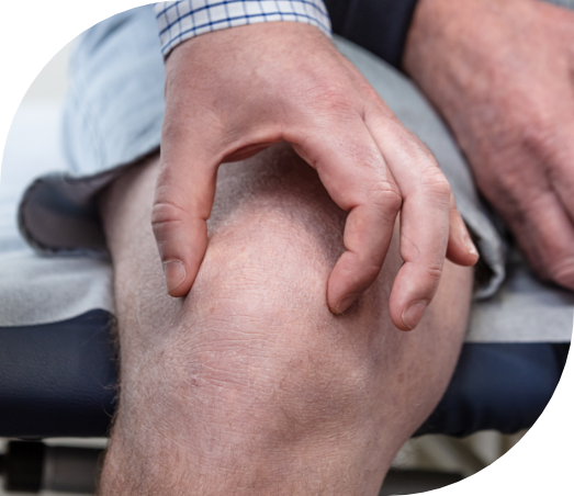
What are the symptoms of a Meniscus tear?
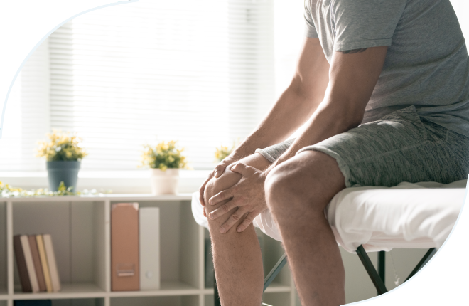
Many people report that they have a ‘torn cartilage’ but in fact the majority actually have a meniscus tear. Most meniscus tears occur from a weight bearing twisting type injury. Some middle aged and older patients may have tears without a specific injury and these are often a result of degeneration of the meniscus tissue. Symptoms of meniscus tears can include pain and swelling of the knee.
If the swelling is severe it may even form a ‘baker’s cyst’ which is a swelling in the back of the knee. Some people experience more mechanical symptoms like clicking, catching and locking in the knee.
Treatment options available
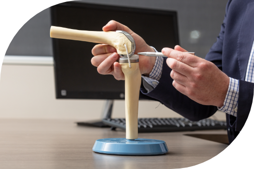
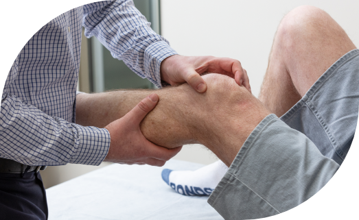
There are various factors that determine the treatment approach. Not all tears of the meniscus are the same. The first step is to make the diagnosis and determine if the tear is the source of pain. This requires a careful history, examination and usually an MRI scan. Chronic tears and tears that occur in older patients generally have less potential to heal. The next step is to determine the type of tear and location of the tear. This is important because only the outer one third of the meniscus receives enough blood flow to provide healing potential. The inner aspect survives on nutrients from the fluid in the knee which limits the repair capabilities. There are various patterns or types of tears that can be seen on MRI.
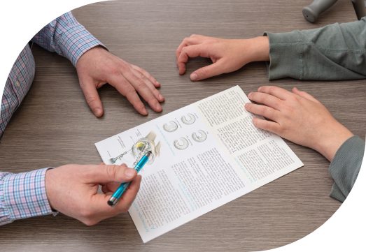
Treatment decisions depend on the size and location of the tear, as well as age and how long the tear has been present. Meniscal tears that occur in younger patients in the peripheral part of the meniscus are often repairable. This involves arthroscopic surgery with a camera in the knee joint to guide carefully placed sutures across the tear to stabilise it. Repairs are best done early after the initial injury. More commonly, tears occur in middle aged patients when the tissue has become more degenerative. Whilst these tears rarely heal, they quite often settle with non-operative treatment. This includes basic rest, ice, elevation and simple analgesics. A good physiotherapy program will help you to regain range of motion, reduce swelling and increase muscle strength around the knee. Cortisone injections may also provide temporary relief. If these conservative measures fail then arthroscopic treatment should be considered to either repair or resect the proud edges of the meniscus.
Dr Woodbridge aims to repair the meniscus where possible. A successful repair has the advantage of reduced contact pressures in the knee joint and likely reduced rates of osteoarthritis in the future. New arthroscopic suturing devices are now available to aid repair techniques. This allows the surgeon repair some of the tear patterns that were previously thought to be unrepairable.
Rehabilitation
Partial Medial Menisectomy
Patients are can be discharged home from the hospital on the same day as the surgery. You will be able to weight bear on the operated leg. Some patients may require crutches to assist their mobbility. It is important to elevate the knee to limit swelling in the knee and lower leg. External bandages should removed after 24 hours, leaving the sterile dressings in place. It is important to perform regular straight leg raise exercises to maintain strength and tone in your quadriceps muscle. Dr Woodbridge will see you approximately 2 weeks after the surgery.
Most patients are quite mobile at this point, but your knee function will continue to improve over approximately 6 weeks. Some patients will benefit from the guidance of a physiotherapist.
Meniscus Repair
Patients are discharged home from the hospital on the same day as surgery. Many patients will need to restrict weight bearing using crutches, and this will assessed by Dr Woodbridge on a case-by-case basis depending on the pattern of the tear. Some patients will have a hinged knee brace. External bandages should be removed after 24 hours, leaving sterile dressings in place. It is important to elevate the knee to limit swelling and to perform regular straight leg raise exercises to maintain strength and tone in the quadriceps muscle.