Chondral Injuries
What is Chondral Tissue?
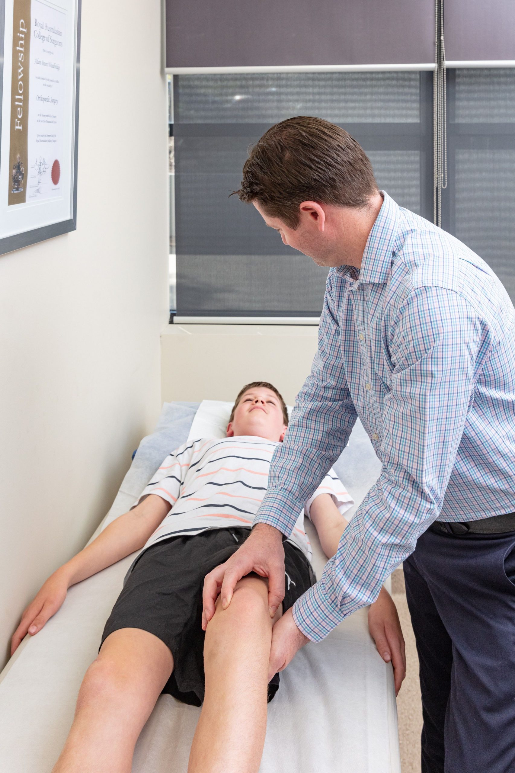
The knee joint contains both the tibio-femoral joint, where the femur (Thigh bone) meets the tibia (leg bone), and the patellofemoral joint where the patella (knee-cap) glides in a groove in the front of the femur. The bone ends or joint surfaces are covered by a smooth layer of articular cartilage or chondral tissue. Cartilage allows almost friction-free and painless motion to occur in the joint.
When defects occur in the cartilage layer, this exposes bone within the joint, creating pain and friction within the joint. Unfortunately, articular cartilage is one of the human tissues that does not tend to re-grow or regenerate when it is worn or injured. A focal chondral injury changes the congruence and force distribution across the joint, which encourages further wear to occur in the surrounding uninjured cartilage. More diffuse wear of the cartilage leads to osteoarthritis of the joint, often at a relatively young age.
Causes of Chondral injuries
Chondral lesions of the knee are most commonly caused by a traumatic injury, either from a direct impact to the knee or from a twisting-type injury that creates a shear-force across the cartilage. Chondral injuries can also be associated with a tear of the meniscus or rupture of ligaments of the knee. Less commonly, cartilage defects may occur when blood flow to the underlying bone is disrupted, a condition called osteonecrosis. The developing bones of children and adolescents are also susceptible to developing focal osteochondral lesions, in a condition called osteochondritis dissecans.
The severity of chondral lesions can be assessed by the depth of cartilage disruption, ranging from mild superficial wear (grade 1) to full-thickness lesions with exposed bone (grade 4). The total surface area of the lesion and the location within the joint also contribute to outcomes. The best form of imaging for a chondral defect is Magnetic Resonance Imaging (MRI). Apart from the size, depth and location, MRI helps to identify if the lesion is associated with a chondral flap, an osteochondral (bone and cartilage) component or even a loose body. Chondral lesions may be apparent in acute presentations (following trauma) or may be chronic (delayed presentation or degeneration).
Surgical options and Treatments
A number of surgical procedures have been developed to manage chondral lesions. Microfracture is a procedure that involves creation of tiny fractures into the exposed bone surface at evenly spaced intervals. This stimulates the body to produce fibrocartilage to fill the defect. Whilst fibrocartilage does not have the same mechanical properties as articular cartilage, it is certainly superior to exposed bone. Autologous chondrocyte implantation is another option but it is not currently funded by medicare and private health funds as the results are similar to those of microfracture.
If the lesion involves an unstable fragment containing both bone and cartilage, then it may be possible to fix this piece back into the defect. Chronic loose bodies may simply require removal of the loose body to prevent locking of the joint and future wear. Patients with abnormal alignment may require an osteotomy to realign the bone, effectively unloading the chondral lesion. Older patients with more diffuse wear may be best served with knee replacement.